Executive Summary
Imaging volumes in radiology are growing faster than healthcare systems can manage, while staffing levels continue to lag. About 69% of radiologists surveyed by the Radiology Business Management Association and American College of Radiology [1] reported being understaffed. At the same time, the demand for imaging continues to rise across emergency, oncology, and preventive screening programs. The result: persistent backlogs, delayed reporting, and increasing clinician burnout.
Artificial intelligence (AI) in healthcare imaging is now emerging as the most practical lever to close this gap. Between 1995 and 2024, the U.S. FDA cleared more than 1,000 AI/ML-enabled medical devices, of which 621 (84.4%) relied on medical imaging as their core input. Notably, radiology served as the lead review panel for most of these approvals (88.2%) [2], underscoring how central imaging has become to AI innovation in healthcare.
These AI-driven medical imaging systems enhance four strategic domains: automated image analysis, early disease detection, predictive insights, and remote diagnostics. Together, these pillars help hospitals shorten turnaround times, reduce reporting variation, and extend the reach of diagnostic imaging automation into underserved regions. However, implementing these AI capabilities requires robust engineering infrastructure and seamless integration with existing radiology systems—a technical challenge that demands specialized software development expertise.
Matellio, as a custom software development and engineering company, bridges this gap by helping healthcare organizations build and integrate AI functionality into their medical imaging platforms. Through tailored engineering services—including PACS/RIS integration, API development, and cloud infrastructure—Matellio enables the technical transformation needed to operationalize AI in diagnostic radiology while maintaining HIPAA and GDPR compliance.
In this article, we will examine how healthcare providers are operationalizing AI across these pillars, and how the technology is integrated into radiology workflows to deliver clinical precision and operational agility.
I. Market Pressure and Imaging Bottlenecks in Healthcare
Medical data visualization and imaging have become both the foundation and the bottleneck of modern healthcare. Radiology departments face surging scan volumes driven by aging populations, wider insurance coverage, and greater clinical reliance on cross-sectional imaging across specialties, from oncology to orthopedics.
Each year, an estimated 4 billion [3] imaging procedures are performed globally, covering CT, MRI, X-ray, and ultrasound. Yet the systems that capture, store, and interpret this volume of data are under severe strain. In many U.S. hospital radiology departments, scan backlog, staff shortages, and delayed reporting have become the norm.
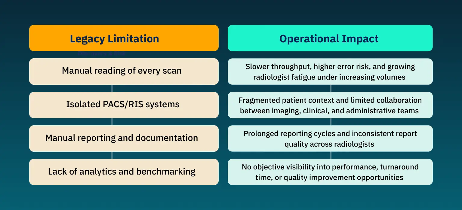
Why Traditional Imaging Workflows Are Reaching Their Limits
For many years, imaging interpretation followed a linear and manual process: acquire, store, read, and report. Many of them were developed two decades ago and relied on manual interpretation, separate ACS/RIS systems, repetitive documentation, and limited analytics.
They falter when faced with high volumes of imaging data, evolving regulatory demands, and the need for intelligent workflow automation. Their key limitations and operational impacts are:
Hospitals today require a new layer of intelligence that integrates imaging data, clinical information, and workflow automation within a single, compliant architecture.
II. How AI is Redefining Diagnostic Precision and Radiology Efficiency
Radiologists are expected to interpret an ever-growing number of scans with the same or fewer resources, while hospitals face pressure to maintain accuracy, speed, and regulatory compliance. The result is an environment where even minor inefficiencies compound into diagnostic delays and clinician fatigue.
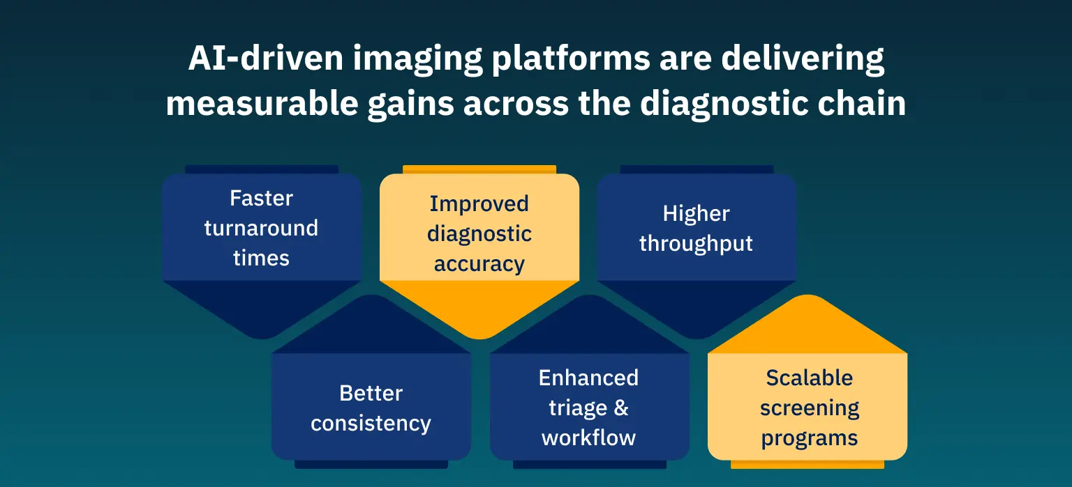
Artificial intelligence is changing the situation. Instead of relying on manual interpretation and linear workflows, AI integration in hospitals now automates repetitive and time-intensive tasks such as image triage, automated anomaly detection, and quantitative measurement. This allows radiologists to focus on higher-value interpretation and clinical decision-making.
A peer-reviewed study found that radiologists using a radiology-specific AI model for chest radiograph interpretation reduced their average reading time by approximately 25% (from 25.8 seconds to 19.3 seconds) per case [4]. Similarly, in CT pulmonary embolism studies, AI-based triage software significantly shortened report turnaround times by prioritizing critical findings for immediate review.
III. Components and Clinical Impact of the AI-Powered Imaging Ecosystem
Modern AI medical imaging software brings together four components that function as a cohesive system:
| Component | What it does | Why it matters |
|---|---|---|
| Data ingestion and preprocessing | Normalizes DICOM inputs across modalities and scanners | Ensures the model sees consistent data, enabling robust inference |
| Model inference engines | Detect pathologies, segment anatomy, and quantify biomarkers | Automates tasks that used to require manual effort |
| Workflow orchestration | Prioritizes urgent cases, routes results, and manages dashboards | Integrates AI into radiologist workflows, not just as a separate tool |
| Continuous learning and feedback | Incorporates new labels, adapts to new scanners/patient demographics | Keeps accuracy high over time rather than degrading |
Together, these systems provide a second set of eyes, a faster analytical layer, and a foundation for predictive and preventive care. The rapid adoption of such technologies reflects their tangible impact: market analysis estimates the global artificial intelligence in diagnostics and medical imaging segment at USD 1.36 billion, with projections to surpass USD 19.78 billion by 2033 [5], growing at a CAGR of 34.67%.
In practice, AI in healthcare imaging helps in:
Automated Image Analysis
Image analysis automation is among the most mature applications of AI in radiology. Deep convolutional neural networks interpret CT, MRI, and ultrasound scans with high consistency, analyzing pixel intensity and spatial relationships faster than the human eye.
- For U.S. hospitals, this translates into measurable efficiency. Research shows that AI reduced turnaround time for cervical spine CT fracture reports from 225.7 minutes to 126.7 minutes [6], a 56.1% improvement.
- Image segmentation algorithms can also generate structured quantitative imaging biomarkers (tumor volume, perfusion rates, and vessel stenosis) that improve reproducibility across radiologists and over time.
- Segmentation algorithms also produce quantitative imaging biomarkers such as tumor volume, perfusion rate, and vessel stenosis, enhancing reproducibility across readers and over time.
Operationally, automation eliminates repetitive measurements, allowing radiologists to focus on complex pathologies and treatment planning. Integrated into PACS viewers, clinical decision support with AI appears as overlayed annotations for quick verification.
2.Early Disease Detection and Scalable Screening
Early detection makes all the difference between reactive and preventive medicine. AI is now improving screening program sensitivity, particularly in oncology, cardiology, and neurology.
- In breast cancer programs across Europe and the United States, AI-assisted mammography has shown 26.4% higher detection rates [7] than those of GRs without AI-CAD.
- Similarly, stroke triage systems powered by AI have cut the time from scan to intervention, significantly improving outcomes in ischemic cases.
This capability not only saves lives but also reduces long-term treatment costs in value-based reimbursement models now prevalent across U.S. healthcare systems.
3.Predictive Insights and Intelligent Treatment Planning
As hospitals digitize imaging archives, they accumulate longitudinal data that reveal how disease evolves. AI algorithms trained on such datasets can recognize subtle progression patterns and forecast likely outcomes.
- For oncologists, predictive imaging technology yields dynamic tumor-response models that indicate when to adjust chemotherapy regimens.
- Cardiologists use similar tools to predict the likelihood of heart-failure readmission by linking MRI biomarkers with electronic health record metrics.
- Hospitals implementing predictive imaging technology report both improved resource allocation and tighter integration between diagnostic and therapeutic departments.
However, successful adoption demands transparency. The rise of explainable AI (XAI) ensures that models provide visual or textual reasoning, helping build trust with radiologists and regulators alike.
4.Remote Diagnostics and the Rise of Distributed Care
Healthcare is shifting toward distributed networks: urgent-care centers, rural clinics, and mobile imaging units. This requires the same diagnostic precision as tertiary clinics. AI integration in hospitals makes this possible through remote diagnostics and tele-radiology ecosystems.
- Cloud-native imaging platforms, combined with edge inference technology, enable scans taken at community sites to be analyzed instantly. Only flagged studies are escalated for specialist review, reducing bandwidth and turnaround time.
- Federated learning, another emerging method, enables institutions to train AI models collaboratively without exchanging patient data, thereby meeting both HIPAA and GDPR standards. This framework protects privacy while creating more robust, generalized algorithms suitable for multi-institution deployment.
IV. How Matellio Enables AI Transformation in Medical Imaging
Even as the benefits of AI-enabled image processing grow clearer, challenges persist. Many institutions begin with pilot programs that never scale because they underestimate the effort needed for annotation, regulatory submission, and workflow alignment.
Matellio collaborates with hospitals, diagnostic networks, and medtech enterprises to turn promising concepts into scalable AI healthcare solutions. By combining engineering precision with healthcare domain expertise, Matellio helps organizations enhance imaging intelligence and clinical decision support with AI integration.
The following case illustrates how a leading medtech company worked with Matellio to strengthen its imaging platform and deliver an investor-ready proof of concept under tight timelines.
Accelerating Proof of Concept for 7D Imaging
Challenges
The 7D Imaging team needed to transform its partially developed mobile application into an investor-ready proof of concept. The existing build lacked the speed, stability, and user experience required to demonstrate the platform’s technical potential.
The team faced tight deadlines, limited engineering resources, and the need to balance rapid execution with architectural integrity.
Solution
Matellio stepped in as a trusted engineering partner to optimize the existing React Native and Expo framework rather than rewriting it from scratch. The team streamlined workflows, enhanced the UI/UX for better usability, and established seamless backend integration on AWS to improve scalability and reliability. This strategic takeover ensured continuous delivery without disrupting prior progress.
Outcomes
- POC delivered ahead of schedule, enabling early investor demos.
- Improved navigation and AWS integration.
- Refined, production-ready prototype boosted stakeholder trust.
- Agile engagement ensured consistent communication and alignment.
V. The Future of Artificial Intelligence in Diagnostics and Radiology
Artificial intelligence in diagnostics has redefined medical imaging from a support tool into a strategic engine for predictive, precise, and patient-centered care. Hospitals no longer view imaging as a passive record but as a continuous source of intelligence that shapes therapy design, staffing efficiency, and patient outcomes. As AI systems mature, their influence extends beyond radiology, informing oncology treatment paths, surgical planning, and population health analytics.
Over 70% of radiology departments and almost 60% cancer departments are already using AI tools today [8]. It shows how hospitals that integrate explainable, interoperable AI platforms into their imaging ecosystems will define the next decade of healthcare precision, cost control, and trust.
Key Takeaways
- AI-driven automation is essential to manage growing imaging volumes and staffing gaps.
- Automated analysis, early detection, and predictive insights are redefining imaging workflows.
- Secure, interoperable, and compliant data pipelines enable scalable AI adoption.
- XAI enhances transparency, fairness, and regulatory trust.
- Integrated data models enable early intervention and personalized therapy planning.
FAQ’s
AI algorithms detect and quantify features invisible to the human eye, helping radiologists identify abnormalities with greater precision. Many studies show significant improvements in sensitivity and reductions in false negatives across CT and MRI modalities.
Hospitals face data-quality issues, workflow integration hurdles, regulatory complexity, and clinician adoption barriers. Each can be mitigated through strong governance and iterative deployment.
AI identifies micro-level tissue changes or perfusion anomalies that signal disease before symptoms appear, supporting large-scale screening and preventive intervention.
AI serves as a decision-support layer, while responsibility for diagnostic imaging automation and contextual judgment remains with humans. The best outcomes occur when both work in tandem.
Transparency, fairness, and data privacy must guide model design and deployment. Explainable AI and bias auditing ensure accountability and public trust.
References:
[1] Radiology Business, Understaffing is ubiquitous in radiology, according to the new ACR/RBMA workforce survey
[2] Nature, How AI is used in FDA-authorized medical devices: a taxonomy across 1,016 authorizations
[3] World Health Expo, Rise of mobile medical imaging to revolutionise healthcare
[5] Grand View Research, AI In Medical Imaging Market
[6] Auntminnie, AI reduces time to interpretation of CT cervical spine exams
[8] The Royal College of Radiologists, The Future of AI in Healthcare: Public perceptions of AI in Radiology